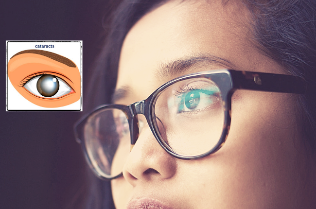
A cataract can be described as a painless clouding of the lens in the eye which can cause decreased vision. Cataracts prevents light from entering the eye, which causes a decrease in image quality.
Most cataracts are age-related. An American study found that more than half of all Americans over the age of 80 either have a cataract or have had cataract surgery. A cataract cannot spread from one eye to the other, but they can present monocularly (in one eye only) or binocularly. Women are more likely to develop cataracts than men.
How do cataracts form?
Cataracts form in the lens of the eye, which is located behind the iris The lens focuses light that passes through the eye on the retina – the layer of nerve tissue lining the eye’s rear and innermost potion and functions like the film of a camera. Cataracts scatter light that passes through the eye which causes images to appear hazy.
With age the lens loses elasticity and become less transparent. Collagen tissues within the lens tend to coagulate and clump together causing non-transparent areas. As the cataract progress it becomes denser and has a greater impact on vision.|
Cataracts tend to develop in both eyes and are usually not symmetrical – becoming more advanced in one eye than the other.
Types of cataracts
Cataracts may develop at birth or may be secondary in nature, developing as a result of diseases such as myopia, uveitis, and hereditary fundus dystrophies. Trauma, infrared radiation, and ionizing radiation may also cause unilateral cataracts. Cortical, nuclear, and posterior subscapular forms develop due to advancing age.
What causes cataracts?

The exact cause of cataracts is unknown, however increased risk factors include the following:
- Advanced Age
- Diabetes
- Smoking
- Eye trauma
- Excessive use of alcohol
- Prolonged use of corticosteroids
- Prolonged exposure to sunlight or radiation
- Nutritional deficiency
- High blood pressure
- Previous eye injury or inflammation
- Previous eye surgery
Signs & Symptoms
Depending on the severity and progression of the cataract – symptoms may include:
- Blurry, foggy or cloudy vision
- Sudden change in prescription
- Colour vision changes as the discoloured lens acts as a filter
- Increased sensitivity to glare from light especially at night
- Double vision or ghost images
- Halos around lights
How are cataracts diagnosed?
- Patient history
Optometrists take a comprehensive patient history to determine the visual difficulties experienced by the patient during his/her daily activities and to get a summary of the patient’s overall health.
- Visual acuity measurement and refraction
Visual acuity and refraction are measured to determine if a sudden change in prescription is present and to what extend the cataract is affecting the vision at distance and near.
- Evaluation of the lens
Under high magnification and illumination, the optometrist examines the lens to determine the extent and location of the cataract.
Once the optometrist is convinced of the presence and severity of the cataract – the patient is referred to an eye specialist (ophthalmologist) for further evaluation and to determine the appropriate treatment plan, which usually entails surgical removal of the opacified lens, followed by an intraocular lens transplant.
Cataract surgery types and risk factors
- Phacoemulsification
Phacoemulsification is presently the most popular method of removing cataracts. Minimum sedation is needed during this procedure which usually takes half an hour. Only local anesthesia is administered and no stiches are needed to close the wound, which speeds up healing time and reduces the risk of scarring. The eye is usually sensitive to sunlight immediately post-surgery due to eye drops used to dilate the pupil, but normal vision returns within a few hours. The ophthalmologist will schedule a follow-up appointment approximately four weeks after surgery, to ensure that the lens has settled and that no residual swelling is present. At his time, another refraction will be performed to evaluate if further spectacle correction is necessary.
- Extracapsular Removal of Cataracts
This procedure is mainly used for more advanced cataracts. This procedure differs in that the lens is not fragmented into smaller parts (phacoemulsify) before it is removed, but rather more incisions are made to take the lens out. Stitches are required after the surgery. The recovery of vision is usually slower. With this procedure generally an eye patch might be needed for some time after surgery.
- Intracapsular Cataract Removal
To perform this surgical method an even larger incision is needed than for extracapsular cataract surgery. The whole with the capsule is removed during this procedure. The intraocular lens is required to be placed in a location in front of the iris. This method is not used that often anymore but may be necessary for cases involving severe eye trauma.
 Who is a candidate for cataract surgery?
It may be mentioned by your eye care professionals during a routine eye examination that you present with early stage lens changes, which may not yet cause you to experience any symptoms of visual loss.
Eye surgery is recommended for most individuals who manifest symptoms secondary to lens opacification, which cannot be corrected by prescription or contact lens wear. Eye surgery may not be recommended if other non-related ocular diseases are present. In such cases, smooth contact lenses that lock moisture in may be prescribed for maximum comfort. This is why the ophthalmologist will do a full, comprehensive ocular examination beforehand to determine the most appropriate course of action.
Is it okay for patients with cataracts to wear tinted contact lenses?
Patients with cataracts may wear colored contact lenses with vision correction provided that they are prescribed by a licensed eye care professional. To ensure premium quality, only choose contact lenses from FDA-approved brands.
